


 18 / 116

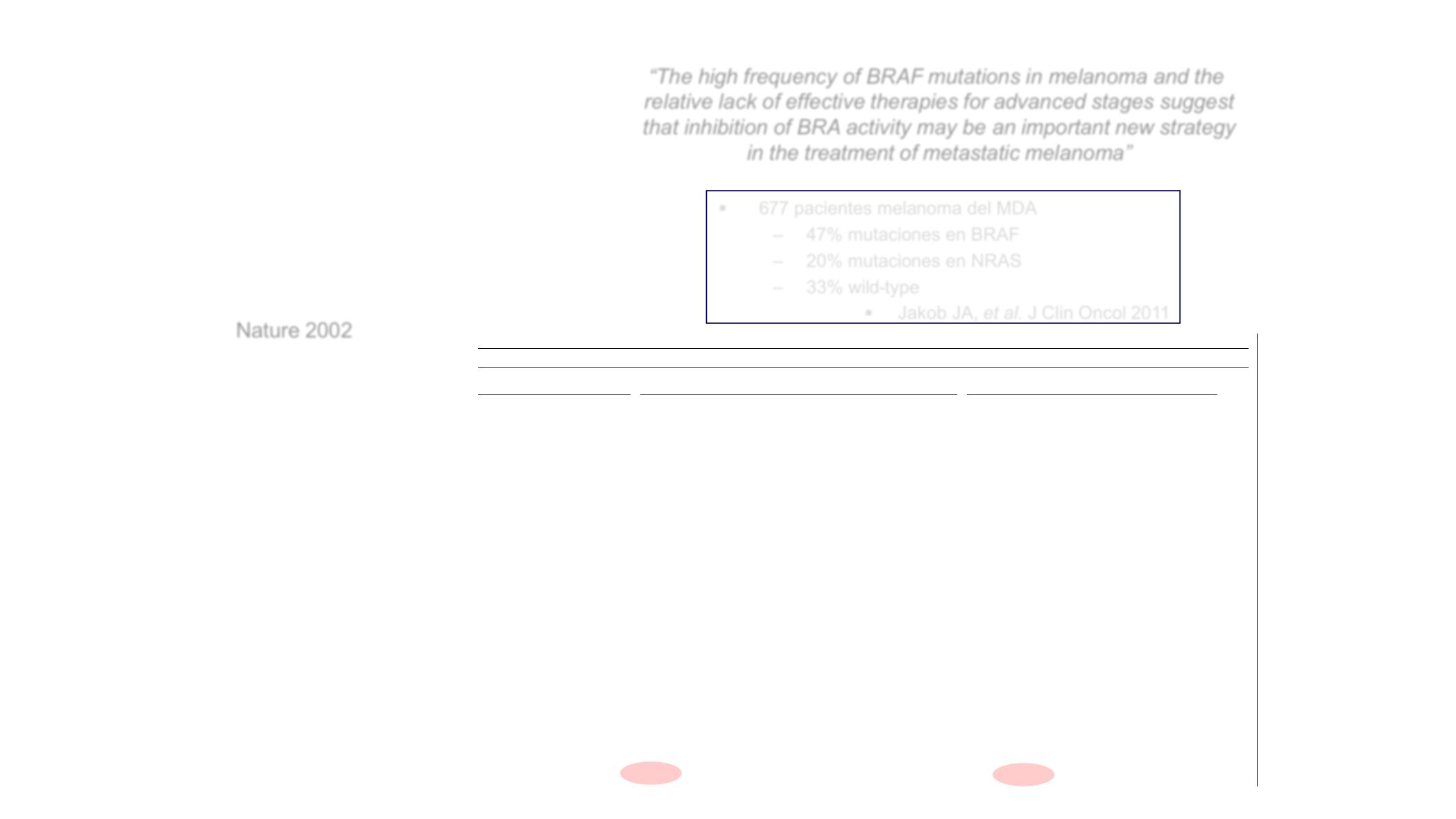
Fig. 1 for example), and 1 of 182 sarcomas. Mutations were not
detected in 33 breast cancers, 15 gliomas, 23 prostate cancers, 14
lung cancers, or 19 head and neck squamous cell carcinomas. Ten of
the 35 ovarian tumours examined were classified as borderline (low
malignant potential) lesions and 4 of 5
BRAF
mutations found in
ovarian neoplasms were in this subcategory. The single primary
sarcoma in which a
BRAF
mutation was found was classified as a
malignant fibrous histiocytoma.
Although
BRAF
mutations are found in a wide range of cancers,
there is a trend towards the occurrence of mutations in cancer types
in which a substantial proportion of cases are known to harbour
RAS
mutations (for example, malignant melanoma, colorectal
cancer and borderline ovarian cancers
4–6
). The apparent association
between the presence of
BRAF
and
RAS
mutations in similar cancer
signalling pathway controlling proliferation and differentiation
operates through activation of
BRAF
and that this gene is mutated
in most melanomas suggests a possible explanation for the high
frequency of
BRAF
mutation in melanomas relative to other cancer
types.
Our analysis reveals mutations in two regions of the
BRAF
kinase
domain. Mutations were very similarly distributed in cancer cell
lines and primary cancers. A total of 89% of mutations are within or
immediately adjacent to the activation segment, a region of 10–30
amino acids bounded by almost invariant DFG and APE motifs
10
.
Acidic substitutions at a single amino acid residue (usually V599E
and one instance of V599D) account for 92% of activation segment
mutations with five further mutations altering residues E585, F594,
G595 and L596 (Table 1). These residues are identical at the
Table 1
BRAF
mutations in human cancer
BRAF
mutations
Cancer cell lines
Primary tumours
(1)
(2)
(3)
(4)
(5)
(6)
(7)
(8)
(1)
(2)
(3)
(4)
(5)
(6)
Nucleotide
Amino acid Mel. Colo. ca. Glioma Lung ca. Sarcoma Breast Ovarian Other Mel. STC Mel. Colo. ca. Ovarian* Sarcoma Other† Total
...................................................................................................................................................................................................................................................................................................................................................................
G1388A
G463E
1
1
G1388T
G463V
1
1
G1394C
G465A
1
1
G1394A
G465E
1
1
G1394T
G465V
1
1
G1403C
G468A
2
2
G1403A
G468E
1
1
G1753A
E585K
1
1
T1782G
F594L
1
1
G1783C
G595R
1
1
C1786G
L596V
1
1
T1787G
L596R
1
1
T1796A
V599E 19
5
4
5
1
1
11
5
2
3
1
0 57
TG1796-97AT
V599D 1
1
Total
20
7
4
4
5
1
1
1
12
6
4
5
1
0 71
No. samples screened
34
40
38
131
59
45
26 172
15
9
33
35
182
104 923
Per cent
59% 18% 11% 3% 9% 2% 4% 0.6% 80% 67% 12% 14% 0.5% 0% 8%
...................................................................................................................................................................................................................................................................................................................................................................
Amino acid residues are grouped in blocks. Three further
BRAF
coding sequence variants were identified (G2041A R681Q in the HEC1A endometrial cancer cell line, T974C I325T in the ZR-75-30
breast cancer cell line, and C2180T A727V in the H33AJ-JA1 T-ALL cell line). These were not present in 341 control DNAs. Lane numbers (in parentheses) are provided for convenience. Mel.,
..............................................................
Mutations of the
BRAF
gene
in human cancer
Helen Davies
1,2
, Graham R. Bignell
1,2
, Charles Cox
1,2
, Philip Stephens
1,2
,
Sarah Edkins
1
, Sheila Clegg
1
, Jon Teague
1
, Hayley Woffendin
1
,
Mathew J. Garnett
3
, William Bottomley
1
, Neil Davis
1
, Ed Dicks
1
,
Rebecca Ewing
1
, Yvonne Floyd
1
, Kristian Gray
1
, Sarah Hall
1
,
Rachel Hawes
1
, Jaime Hughes
1
, Vivian Kosmidou
1
, Andrew Menzies
1
,
Catherine Mould
1
, Adrian Parker
1
, Claire Stevens
1
, Stephen Watt
1
,
Steven Hooper
3
, RebeccaWilson
3
, Hiran Jayatilake
4
, Barry A. Gusterson
5
,
Colin Cooper
6
, Janet Shipley
6
, Darren Hargrave
7
, Katherine
Pritchard-Jones
7
, Norman Maitland
8
, Georgia Chenevix-Trench
9
,
Gregory J. Riggins
10
, Darell D. Bigner
10
, Giuseppe Palmieri
11
,
Antonio Cossu
12
, Adrienne Flanagan
13
, Andrew Nicholson
14
Judy W. C. Ho
15
, Suet Y. Leung
16
, Siu T. Yuen
16
, Barbara L. Weber
17
,
Hilliard F. Seigler
18
, Timothy L. Darrow
18
, Hugh Paterson
3
,
Richard Marais
3
, Christopher J. Marshall
3
, Richard Wooster
1,6
,
Michael R. Stratton
1,4
& P. Andrew Futreal
1
1
Cancer Genome Project, The Wellcome Trust Sanger Institute, Wellcome Trust
Genome Campus, Hinxton, CB10 1SA, UK
3
Cancer Research UKCentre for Cell and Molecular Biology, Chester Be tty Labs,
Institute of Cancer Research, London SW3 6JB, UK
4
Section of Cancer Genetics;
6
Section of Molecular Carcinogenesis; and
7
Section
of Paediatrics, Institute of Cancer Research, Sutton, Surrey SM2 5NG, UK
5
Department of Pathology, Western Infirmary, University of Glasgow, S11 6NT,
UK
8
Department of Biology, YCR Cancer Research Unit, University of York,
York YO10 5YW, UK
9
Queensland Institute of Medical Research, RBH Post Office Herston, Queensland
4029, Australia
10
Department of Pathology, and
18
Department of Surgery, Duke University
Medical Centre, Durham, North Carolina 27710, USA
11
Institute of Molecular Genetics, C.N.R., Loc. Tramariglio, Alghero 07040, Italy
12
Department of Pathology, University of Sassari, Azienda USL1, Sassari 07100,
Italy
13
Royal Free
&
University College Medical School, London WC1E 6JJ, UK
14
Royal Brompton Hospital, London SW3 6NP, UK
15
Department of Surgery, and
16
Department of Pathology, The University of Hong
Kong, Queen Mary Hospital, Hong Kong
17
Abramson Family Cancer Research Institute, University of Pennsylvania Cancer
Center, Philadelphia, Pennsylvania 19104, USA
2
These authors contributed equally to this work
.............................................................................................................................................................................
Cancers arise owing to the accumulation of mutations in critical
genes that alter normal programmes of cell proliferation, differ-
entiation and death. As the first stage of a systematic genome-
wide screen for these genes, we have prioritized for analysis
signalling pathways in which at least one gene is mutated in
human cancer. The RAS–RAF–MEK–ERK–MAP kinase pathway
mediates cellular responses to growth signals
1
. RAS is mutated to
an oncogenic form in about 15% of human cancer. The three
RAF
genes code for cytoplasmic serine/threonine kinases that are
regulated by binding RAS
1–3
. Here we report
BRAF
somatic
missense mutations in 66% of malignant melanomas and at
lower frequency in a wide range of human cancers All mutations
are within the kinase domain, with a single substitution (V599E)
accounting for 80%. Mutated BRAF proteins have elevated kinase
phoblastoid cell lines from the same individuals were screened for
sequence variants through the coding exons and intron–exon
junctions of the
BRAF
gen using a capillary-based modified
heteroduplex method followed by direct sequencing of polymerase
chain reaction products. (Exon 1, containing 135 base pairs (bp) of
coding sequence, failed to amplify despite the use of five different
primer sets.) Three single-base substitutions were detected. T o
were in
BRAF
exon 15: T1796A leading to a substitution of valine by
glutamic acid at position 599 (V599E) in the melanoma cell line
Colo-829, and C1786G leading to L596V n th NSCLC cell line
NCI-H2087 (Fig. 1). A further mutation was found in exon 11:
G1403C leading to G468A in the NSCLC cell line NCI-H1395. None
of the three changes were present in the lymphoblasto d cell lines
from the same individuals, indicating that the variants were soma-
tically acquired mutations.
l tters to n ture
§
677 pacientes melanoma del MDA
– 47% mutacione en BRAF
– 20% mutaciones en NRAS
– 33% wild-type
§
Jakob JA,
et al.
J Clin Oncol 2011
“Th high frequency of BRAF mutations i lanom and the
relative lack of effective therapies for advanced stages suggest
that inhibition of BRA activity may be an important new strategy
in the treatment of metastatic melanoma”
Nature 2002